

Bone health is one of the most overlooked aspects of long-term wellbeing. Unlike weight, blood pressure, or cholesterol, bone density does not give obvious day-to-day signals when it begins to decline. There is no pain in the early stages. No warning light. No visible change.
For many people, the first indication that something is wrong comes far too late, often in the form of a fracture after a simple fall, sudden back pain caused by a vertebral compression injury, or a gradual loss of height that goes unnoticed for years.
This silent nature of bone loss is precisely why DEXA scanning has become the global gold standard for assessing skeletal health. It provides a precise, low-risk measurement of bone mineral density and allows clinicians to identify osteopenia and osteoporosis long before fractures occur.
Yet one question often surprises patients and readers alike. Why does a DEXA scan focus almost exclusively on two areas of the body: the lumbar spine and the hip?
At first glance, it seems limited. However, this targeted approach is not a shortcut. It is a scientifically validated strategy built on decades of research in bone biology, fracture prediction, and clinical outcomes.
To understand why these two regions matter so much, we need to explore how bone behaves, how it weakens, and why not all skeletal sites tell the same story.
Understanding Bone Density and Why It Matters
Bone is living tissue. It is constantly being broken down and rebuilt through a process known as bone remodelling. During childhood and early adulthood, bone formation exceeds bone breakdown. This allows the skeleton to grow in size, strength, and density, reaching what is known as peak bone mass, typically in the late twenties or early thirties.
After this peak, the balance slowly begins to shift. Bone resorption starts to exceed formation. The result is a gradual decline in bone mineral density over time. This process is natural, but its rate varies widely depending on genetics, hormones, nutrition, lifestyle, and medical conditions.
A DEXA scan measures this density using very low-dose X-rays. The result is a highly accurate snapshot of bone mineral content, usually expressed in grams per square centimetre.
From this measurement, clinicians calculate two key values:
T-score, which compares your bone density to a healthy young adult reference population
Z-score, which compares your bone density to that of others of the same age and sex
These scores are used to classify bone health into normal, osteopenia (low bone mass), or osteoporosis (significantly reduced bone density and increased fracture risk).
However, the reliability of these scores depends heavily on where the measurement is taken. That is where the spine and hip become essential.
Why the Lumbar Spine Is One of the First Areas Scanned
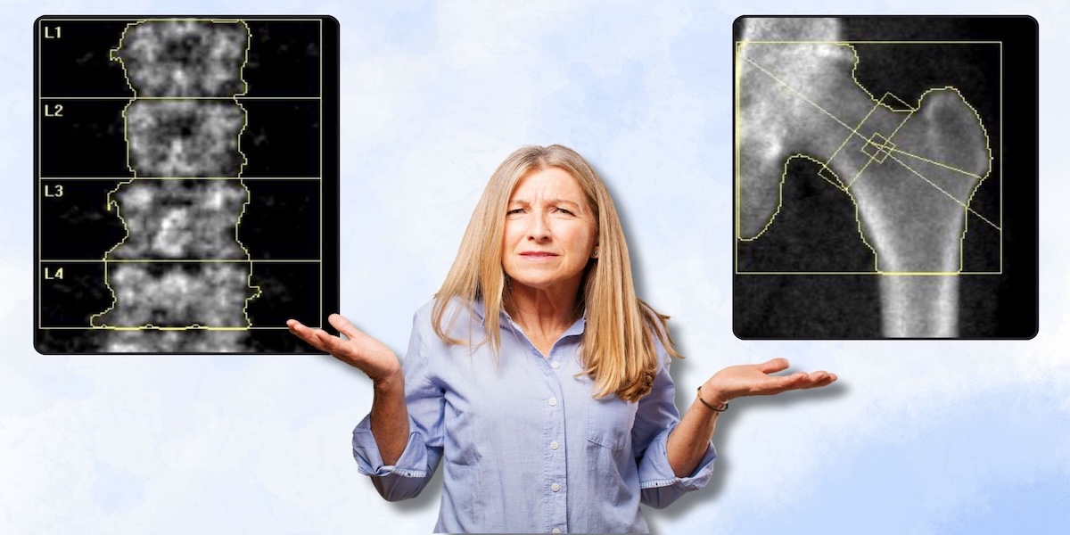
The lumbar spine is located in the lower back and consists of five vertebrae. We scan L1 - L4 for a DEXA Bone Scan. These bones are structurally different from many other parts of the skeleton because they contain a high proportion of trabecular bone.
Trabecular bone, sometimes called spongy bone, has a porous, lattice-like structure. It is highly metabolically active and undergoes rapid turnover compared to cortical bone, which forms the dense outer shell of most bones.
Because of this high metabolic activity, the lumbar spine is extremely sensitive to changes in bone metabolism. When bone loss begins, it often appears here first.
This is particularly important in several scenarios:
Hormonal changes
In women, the decline in oestrogen during menopause can accelerate bone loss significantly. The lumbar spine is often one of the earliest areas to reflect this hormonal shift.
Nutritional and lifestyle factors
Low calcium intake, vitamin D deficiency, low protein consumption, and prolonged calorie restriction can all affect bone turnover. The spine tends to respond quickly to these changes.
Medical conditions and medications
Long-term steroid use, thyroid disorders, and certain chronic conditions can lead to accelerated bone loss that becomes visible early in the spine.
Because of this sensitivity, the lumbar spine is an invaluable early warning system. It detects changes before they become clinically severe, allowing intervention at a stage where lifestyle changes can still make a meaningful difference.
It is also useful for monitoring treatment response. Improvements in bone density are often detectable sooner in the spine than in other skeletal regions due to its high turnover rate.
Why the Hip Is Equally Important
While the lumbar spine is excellent for detecting early changes, the hip plays a very different role in bone health assessment.
The hip is one of the most clinically significant fracture sites in the human body. Hip fractures are associated with serious consequences, including loss of independence, reduced mobility, long-term rehabilitation, and increased mortality risk in older populations.
Because of this, hip bone density is one of the strongest predictors of future fracture risk.
The hip region contains both trabecular and cortical bone. This makes it structurally more balanced and less sensitive to short-term fluctuations than the spine. It also makes it a more stable indicator of long-term fracture risk.
Another important factor is reliability. The hip is less affected by degenerative changes such as osteoarthritis or calcification, which can sometimes artificially elevate spinal readings in older adults. This makes it a more dependable site for long-term assessment.
Hip measurements are also widely used in fracture risk prediction models, such as FRAX, which estimate the likelihood of fractures over a 10-year period. This makes the hip central to clinical decision-making in osteoporosis management.
Why Both the Spine and Hip Are Needed Together
Using both sites provides a far more complete picture of bone health than either measurement alone.
The lumbar spine provides early detection of bone loss.
The hip provides long-term fracture risk prediction.
Together, they offer a balanced assessment of both current bone status and future risk.
In some cases, results may differ between the two sites. This is not unusual. For example:
A patient may show reduced bone density in the spine but normal hip readings, suggesting early-stage bone loss
Alternatively, the hip may show lower density while the spine remains relatively stable, indicating elevated fracture risk
These differences are clinically useful because they help identify patterns of bone loss that would otherwise be missed.
This dual-site approach is recommended by international osteoporosis guidelines and is considered the gold standard in bone health assessment.
How Spine and Hip Measurements Reflect Whole-Body Bone Health
Although bone density is measured at specific sites, bone loss is generally systemic. This means that when bone density decreases, it tends to affect the entire skeleton over time rather than isolated areas.
The spine and hip act as representative sites because they contain different types of bone and respond differently to physiological changes. This allows clinicians to capture a broad picture of skeletal health using a small number of measurements.
Research has shown strong correlations between bone density in the spine and hip and fracture risk in other areas of the body, including the wrist, shoulder, pelvis, and vertebrae.
This is why whole-body scanning is not required for osteoporosis assessment. The spine and hip provide enough information to reliably assess risk.
Interpreting Your DEXA Results
A typical DEXA report will include measurements for both the lumbar spine and hip, along with T-scores and Z-scores.
A T-score above -1.0 is considered normal
A T-score between -1.0 and -2.5 indicates osteopenia
A T-score below -2.5 indicates osteoporosis
Z-scores help determine whether bone density is lower than expected for age and sex, which may indicate underlying medical conditions.
These scores allow clinicians to guide recommendations, which may include lifestyle changes, supplementation, medication, or further investigation.
Who Should Consider a DEXA Scan
Bone density testing is not limited to older adults. Many people can benefit from early assessment, especially if risk factors are present.
These include:
- Menopause or perimenopause
- Family history of osteoporosis
- Low body weight or eating disorders
- High levels of endurance training
- History of stress fractures
- Long-term steroid use
- Hormonal imbalance
- Low vitamin D levels
- Thyroid conditions
- Sedentary lifestyle
Athletes in particular may be at risk of low bone density due to low energy availability or overtraining.
Lifestyle Factors That Influence Bone Strength
Bone health is highly responsive to lifestyle. Positive influences include resistance training, weight-bearing exercise, adequate protein intake, sufficient calcium, and optimal vitamin D levels.
Negative influences include smoking, excessive alcohol consumption, prolonged calorie restriction, inactivity, and untreated hormonal imbalances.
Because the lumbar spine responds quickly to these factors, it often reflects lifestyle changes earlier than other regions.
Why Early Detection Is So Important
Osteoporosis is often referred to as a silent disease because symptoms do not appear until a fracture occurs. By that point, significant bone loss may have already taken place.
Early detection allows for intervention before fractures happen. Many individuals can stabilise or even improve bone density through targeted lifestyle changes and medical support.
Monitoring over time also provides reassurance and helps track progress.
Frequently Asked Questions
Why not scan the whole skeleton?
Because bone loss is systemic, and spine/hip measurements strongly correlate with overall fracture risk.
Is a DEXA scan safe?
Yes. Radiation exposure is extremely low, significantly lower than a standard X-ray.
Can bone density improve?
Yes. With resistance training, nutrition, and appropriate medical support, bone density can stabilise or increase.
How often should scans be repeated?
Typically every 1–2 years depending on risk profile and previous results.
The Gold Standard for Bone Health
The focus on the lumbar spine and hip in DEXA scanning is not a limitation. It is a precision-based strategy supported by decades of clinical evidence.
The lumbar spine reveals early bone loss.
The hip predicts future fracture risk.
Together, they provide a complete and reliable assessment of skeletal health.
This targeted approach allows clinicians to detect problems early, monitor changes over time, and guide prevention strategies that protect long-term mobility and independence.
Bone health is not simply about avoiding fractures. It is about preserving strength, movement, and quality of life. The spine and hip offer the clearest window into that future.